By Lisa Rapaport
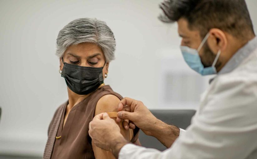
Patients with cancer shouldn’t delay COVID-19 vaccination despite the potential for clinically evident adenopathy on imaging done after vaccination, according to recommendations from a multidisciplinary panel of scientific experts.
Vaccination-associated adenopathy or axillary swelling may appear within two to four days after either vaccine dose and last up to two days with the Moderna vaccine and up to 10 days with the Pfizer-BioNTech vaccine, according to recommendations published in Radiology. This imaging finding may appear when using modalities including mammography, breast MRI, chest and neck CT, and whole-body PET.
To allow time for swelling or adenopathy to resolve prior to imaging, routine cancer screening should be scheduled prior to COVID-19 vaccination or at least six weeks after the final dose, the recommendations state.
But the authors emphasize that clinically urgent imaging for acute symptoms of cancer, short-interval treatment monitoring, planning treatment regimens, or addressing complications should not be delayed based on the timing of any prior vaccination doses. Vaccine doses should be administered on the side of the body contralateral to the location of primary or suspected cancer, and all vaccine doses should be administered in the arm.
“We are observing reactive adenopathy in response to the COVID-19 vaccine in greater numbers than we have with other vaccines, probably due to a combination of the facts that the COVID-19 vaccines elicit a pronounced immune response in recipients and that there is a widescale vaccination effort underway with a large number of people vaccinated in the last two months,” said Dr. Kimberly Feigin, a co-author of the recommendations and acting chief of breast imaging at Memorial Sloan Kettering Cancer Center in New York City.
If radiologists know the dates and sites of the vaccine administration as well as a patient’s cancer history or risk factors, they should in most cases be able to distinguish between adenopathy that is likely a vaccine reaction and adenopathy that is likely malignant, Dr. Feigin said by email. Reactive adenopathy subsides on its own over time, whereas malignant adenopathy does not resolve without treatment.
“In some cases, follow-up imaging may be recommended to ensure that adenopathy subsides when it is believed to be caused by the vaccine,” Dr. Feigin said. “In a minority of cases, needle biopsy of an abnormal lymph node may be recommended to distinguish between benign reactive adenopathy and malignant adenopathy.”
Pre-imaging questionnaires should inquire about any COVID-19 vaccine administration dates, injection sites, laterality and type of vaccine, according to the recommendations.
When imaging does show clinically evident adenopathy after vaccination, patients should wait at least four weeks before a referral for diagnostic imaging evaluation of the nodes, the recommendations also advise.
“Certainly, the Covid-19 vaccine is recommended and should be obtained as the risk of morbidity from the virus outweighs the risk from the vaccine,” said Dr. Gary Cohen, chair and chief of radiology at Temple University Health System in Philadelphia.
“Following simple measures such as waiting 4-6 weeks after completion of their vaccination before obtaining routine or screening mammograms, injection in the opposite arm of known breast cancer, letting the technologist know which arm and when they had their vaccination, and proceeding with diagnostic mammograms if presenting with new breast-related symptoms should both guide timing, patient care, and greatly aid our mammographers in interpretation of these challenging diagnoses,” Dr. Cohen, who wasn’t involved in the recommendations, said by email.
This article was published by Medscape.


