By Peter Hofland, Ph.D
Scientists at the Medical University of South Carolina (MUSC) and the University of California at San Diego (UCSD) are shedding light on a decades-old controversy, showing that autophagy or “self-eating” genes work against tumors in certain types of ovarian cancer. The study was, in part, sponsored by “Nine Girls Ask,” a non-for profit organization focusing on finding a cure for ovarian cancer.
Autophagy is a cellular recycling pathway. Scientists believe that is may play a role in cancer resistance to stresses caused by chemotherapy.
In the study published in PLOS Genetics, the scientists describe that autophagy genes also act to prevent tumor formation.[1]
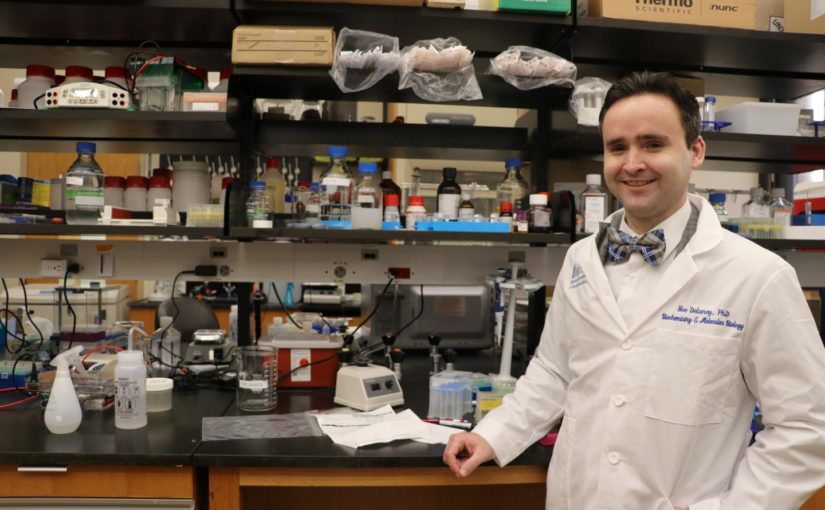
“This finding, validated in mice, helps us to resolve a decades-old controversy based mostly on cells grown in plastic dishes,” explained Joe Delaney, Ph.D., a researcher at Hollings Cancer Center, who was the MUSC faculty lead on the study.
Delaney teamed up with Dwayne Stupack, Ph.D., in the division of gynecologic oncology at UCSD.
Bioinformatic and wet-lab research
Delaney, who uses bioinformatic studies and wet-lab research, studies how aneuploidy, the condition of having an abnormal number of chromosomes in a haploid set,* contributes to disease, particularly in ovarian cancer.
Aneuploidy is the presence of an abnormal number of chromosomes in a cell, which leads to diseases like cancer or conditions like Down syndrome. High-grade serous carcinoma is the most malignant form of ovarian cancer and accounts for up to 70% of all ovarian cancer cases.
BECN1 and LC3B
The researchers focused on two autophagy genes: BECN1 and LC3B. Every person has two copies of these genes: one coming from each of their parents. One copy of BECN1 is lost in one of four breast cancers and three of five serous ovarian cancers. This is unusually frequent for a tumor- suppressor gene.
However, BECN1 has been controversial and complicated. The reason is that this gene is rarely mutated, and even when it is lost, it is often lost with its neighbor, BRCA1, on both human (chromosome 17) and murine (chromosome 11) genomes.
“Some scientists believe that BECN1 deletion is a coincidental event,” Delaney noted.
However, previous studies in cell culture hinted at tumor-initiating characteristics when BECN1 was suppressed, prompting the current study.
“There were strong messages regarding the role of BECN1 in ovarian cancer that hinted that the situation was more complicated. We were working then with a mouse model of ovarian cancer that we could modify at the genetic level. It was time to put the question to rest and measure for tumor suppression directly,” said Dwayne Stupack, Ph.D., in the division of gynecologic oncology at UCSD.
Partial Deletion
To investigate the potential tumor suppressor roles of two of the most commonly deleted autophagy genes in ovarian cancer, BECN1 and MAP1LC3B were knocked-down in atypical (BECN1+/+ and MAP1LC3B+/+) ovarian cancer cells.[1]
Delaney and Stupack performed the same partial deletion found in patients, one of two BECN1 alleles in mammals, in this mouse model. The results were immediate and conclusive. The very first BECN1-suppressed mouse enrolled in the study had a sizeable tumor at 3 months of age.
“Tumors with the model would not normally form until 4 to 6 months. We knew we had discovered something right away,” Delaney said.
Years of research then went into the investigation of why BECN1 single-allele deletion acted as a tumor suppressor. Using the mouse tumors and human ovarian cancer cells within the lab, the team of 15 researchers tested multiple phenotypes to determine why autophagy gene deletion leads to cancer.
The study’s authors suspected cellular metabolism to be one of the strongest effects following the disruption of this recycling pathway. Ultra-performance liquid chromatography mass-spectrometry metabolomics was performed on human ovarian cancer cells with reduced autophagy genes. Surprisingly, little metabolism was changed. Instead, ovarian cancer cells seemed to delete BECN1 to increase the tumor’s ability to evolve.
“Cancer has always been hard to fight because of its ability to evolve in response to treatment. It was clear that removing these genes increased the rate of genetic evolution. Worse yet, many of these changes were random, allowing for the cancer to potentially evolve many different types of resistance,” Delaney explained.
Frightening
While this tumor-suppressor role of autophagy genes is frightening, given how many patients have lost an allele of either autophagy gene, the tumor’s reliance on low autophagy may actually enable the next generation of treatment for ovarian cancer, he said.
“We knew for a long time that autophagy was very different in ovarian cancer. Normal cells in the fallopian tube and on the ovary express high levels of autophagy proteins. Yet dozens of autophagy genes are found to be deleted in a single tumor. This lack of expression helps the tumor form but may also create a weakness for new drugs to target,” Stupack agreed.
“Before this tumor-suppression study, we had already shown that further clogging the cells’ recycling system killed more advanced forms of serous ovarian cancer. Now we understand why the tumors would have this vulnerability in the first place,” he added.
In their prior study, the authors found that autophagy drugs killed the most difficult form of ovarian cancer. They used tumor cells from a cancer patient that were grown in mice.
“Chloroquine, which targets autophagy, has been tried in cancer trials. Ironically, it was used in tumors with very high autophagy levels, the exact opposite of ovarian cancer. We think that, in combination with other autophagy disruptors, chloroquine may have its best effect in a serous ovarian cancer trial because these cells are already compromised. This has yet to be attempted,” Delaney said.
In the article published in PLOS Genetics Delaney and Stupack provide further rationale for why autophagy-targeting drugs should be tested in clinical trials.
Given that multiple autophagy gene losses are present in nine of 10 serous ovarian cancer patients, Delaney hopes to see more research in this area. Ovarian cancer ranks fifth in cancer deaths among women, accounting for more deaths than any other cancer of the female reproductive system.
“Clinical trials are expensive, and these drugs are not profit-generating because they are inexpensive and usually off-patent. We will continue to work to convince the cancer research community autophagy disruption is a worthwhile strategy. We hope to save lives.”
* The presence of extra chromosomes, or missing chromosomes, is a condition called aneuploidy. The risk of having a child with an aneuploidy increases as a woman ages. For example, a human cell having 45 or 47 chromosomes instead of the usual 46. It does not include a difference of one or more complete sets of chromosomes. A cell with any number of complete chromosome sets is called a euploid cell. Trisomy is the most common aneuploidy. In trisomy, there is an extra chromosome. A common trisomy is trisomy 21 (Down syndrome). Other trisomies include trisomy 13 (Patau syndrome) and trisomy 18 (Edwards syndrome). Monosomy is another type of aneuploidy in which there is a missing chromosome. A common monosomy is Turner syndrome, in which a female has a missing or damaged X chromosome.
References
[1] Delaney JR, Patel CB, Bapat J, et al. Autophagy gene haploinsufficiency drives chromosome instability, increases migration, and promotes early ovarian tumors. PLoS Genet. 2020;16(1):e1008558. Published 2020 Jan 10. doi:10.1371/journal.pgen.1008558 [Pubmed]
This article was published by Onco’Zine.


